
"Lameness Imaging Like You've Never Seen It Before"
Led by Dr. Shannon Holmes, UGA CVM Department of Diagnostic Imaging
Bullet points, because this lady had a lot of information and it went so over my head. Also, she had an intelligent, dry wit and made me think of Daria.


- If treatment isn't working, then you have the wrong diagnosis.
- Diagnostic analgesia (blocking): lesions are clinically significant if they correspond with blocking. (Which, I think also can be taken the other way: if there's a lesion but lameness doesn't improve with blocking of that lesion, then it isn't clinically significant.)
- Imaging modalities:
- radiographs (x-rays): due to cost/size/ability to transport, this is still the first line.
- Always take all four directions, because it's easy to miss stuff. In fact, you still might be in the wrong area or performing the wrong test (which leads back to performing correct diagnostic analgesia).
- There has to be a 30-50% increase in bone mineral before the human eye can detect it. So, you know, shit has to be pretty fucked up.
- ultrasound: high user dependency, which means that if the person performing the u/s isn't the best at his or her job, you may miss stuff or misread stuff.
- nuclear scintigraphy: (I'd never heard of this one until I googled it later and found out that this is the fancy name for a bone scan) sensitive, but not specific. Difficult for soft tissue, but good for the sacro-illiac joint. Rood and Riddle has some pretty good information and photos on it.
- computed tomography: AKA, "Cat Scan". She didn't even touch on this one because she said that it just doesn't happen.
- magnetic resonance imaging (MRI): this was Dr. Holmes' favorite. She said that in the past, there was an issue with horses waking up from general anesthesia, but that it is now very safe. She talked a little bit about the two types of MRI magnets and what they were for and showed a few photos, but essentially: it's safe and they're going to shove as much of the horse into the machine as they safely can.
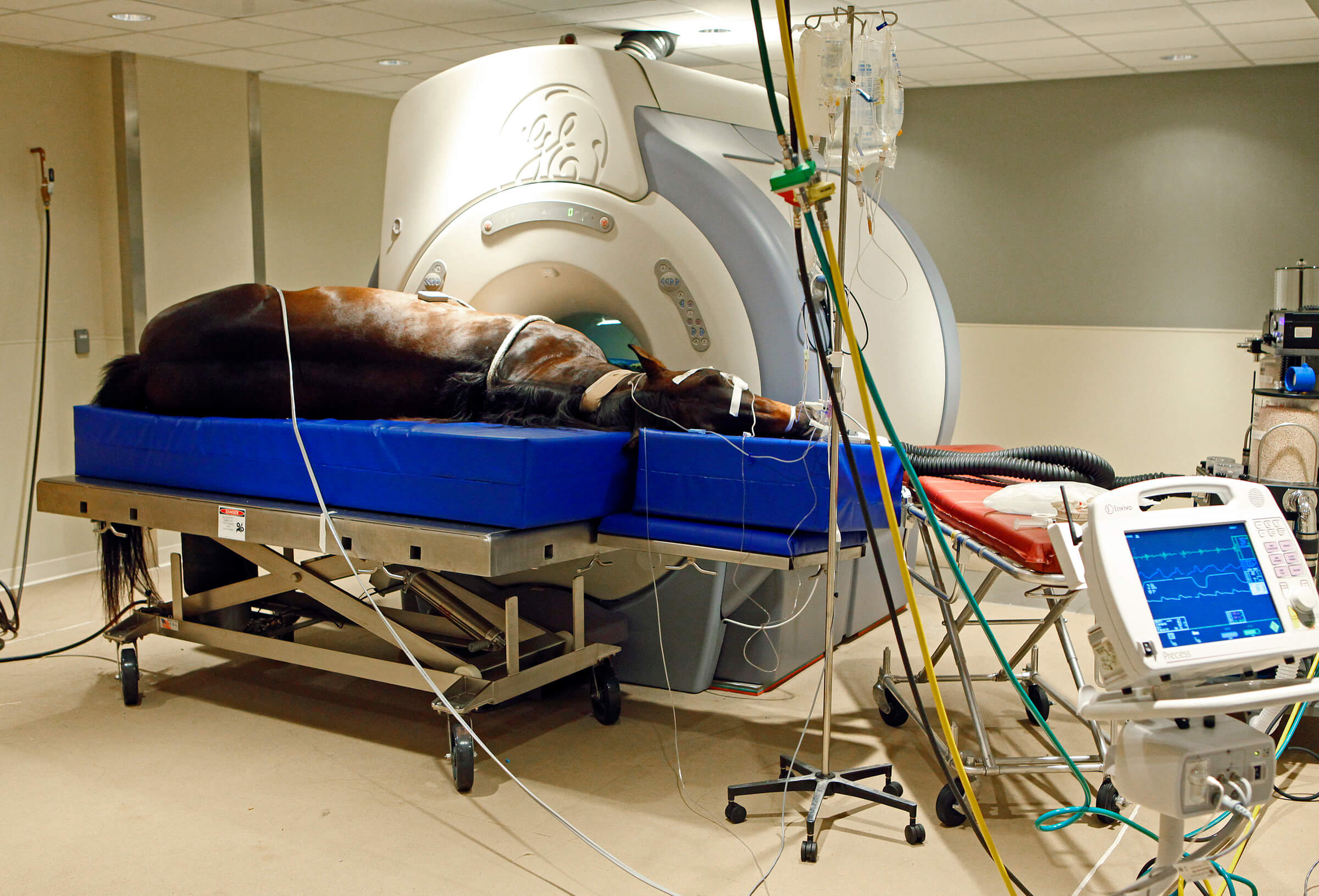 |
| Here's a photo of an MRI. Of course, it isn't a UGA photo because they haven't shared the presentations. (I've requested!) |


.JPG)

6 comments
How do you get the horse on the bed!?
ReplyDeleteA crane - type thing I think...but don't quote me as I'm no vet or any way medically qualified
Delete"made me think of Daria" ahahahahahahaha!!! I loved Daria...
ReplyDeleteVery interesting!
ReplyDeleteGreat information and thank you for sharing! Another big thing nowadays is equine thermography. It's great for locating injuries higher up the leg in more densely muscled areas (like groin and chest) where radiographs and ultrasound can be inconclusive.
ReplyDeletehttp://www.sciencedaily.com/releases/2013/03/130327092521.htm
You have a really awesome vet right there in GA, Dr. Kenneth Marcella, who has been doing some pretty ground-breaking studies on this subject, specifically where it relates to diagnosis of groin and chest injuries, which are a lot more common than originally thought. I saw one of his presentations at the AERC Conference in Atlanta back in March and really enjoyed it. This is one of his articles on the subject:
http://veterinarynews.dvm360.com/equine-adductor-muscle-injuries?id=&pageID=1&sk=&date=
I thought it was really cool. It's just another tool in the box for our modern equine vets and one that can be very useful for diagnosis + can be more affordable than some of the other imaging options for horses that are hard to diagnose. Granted it doesn't provide an image of the injury like the MRI does, but it can help further pinpoint the source of pain on the horse's leg.
that photo of the horse getting an mri is wild! lots of good info tho
ReplyDeleteThanks!